![]() Figure 1 of
Liao, Mol Vis 2004;
10:1038-1046.
Figure 1 of
Liao, Mol Vis 2004;
10:1038-1046.
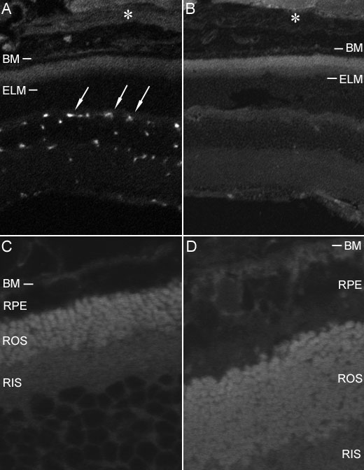
Figure 1. Immunofluorescent localization of albumin in the retina of wildtype mice by laser scanning confocal microscopy
A,C: Sections treated with FITC conjugated rabbit IgG directed against mouse albumin. B,D: Sections treated with antibody preadsorbed with mouse albumin. Note that immunospecific fluorescence was restricted to blood vessels (arrows) and sclera (asterisk). Original magnification in A and B was 20x; magnification in C and D was 189x. The retinal pigment epithelium (RPE), external limiting membrane (ELM), Bruch's membrane (BM), rod outer segments (ROS), and rod inner segments (RIS) are labeled.