![]() Figure 5 of
Davies, Mol Vis 2004;
10:1028-1037.
Figure 5 of
Davies, Mol Vis 2004;
10:1028-1037.
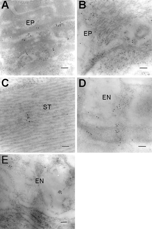
Figure 5. Electron microscopic labelling for ClC-3 in the cornea
A,B: Corneal epithelium. C: Corneal stroma. D-E: Corneal endothelium. Immunolabelling was present in all cell types. Note the tendency for gold particles to appear in clusters. The epithelium (EP), stroma (ST), endothelium (EN) are labeled. All scale bars represent 100 nm.