![]() Figure 4 of
Davies, Mol Vis 2004;
10:1028-1037.
Figure 4 of
Davies, Mol Vis 2004;
10:1028-1037.
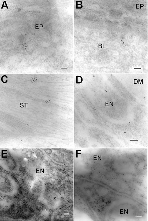
Figure 4. Electron microscopic labelling for ClC-2 in the rat cornea
A: Corneal epithelium. B: Sub-epithelial stroma. C: Middle stroma. D-F: Corneal endothelium. Examples of positive labelling are shown in A-E. Note that gold particles are localized mostly adjacent to plasma membranes, but also intracellularly. A control section (F) is shown for comparison. No gold particles are visible after omission of the primary antibody. The epithelium (EP), Bowman's layer (BL), stroma (ST), endothelium (EN), Descemet's membrane (DM) are labeled. Scale bars represent 100 nm.