![]() Figure 3 of
Davies, Mol Vis 2004;
10:1028-1037.
Figure 3 of
Davies, Mol Vis 2004;
10:1028-1037.
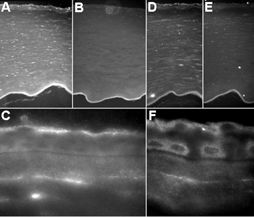
Figure 3. Light microscopic immunofluorescence staining for ClC-2 and ClC-3 in human cornea
A,D: Low power view showing labelling of whole cornea. Note that in A, the endothelial cell monolayer has become detached from Descemet's layer, while in D, the endothelial cell layer is mostly absent. C,F: At higher magnification, some regions show pronounced immunolabelling along the apical region of the superficial epithelial cell layer, and in Bowman's layer. Note also the suggestion of intracellular labelling for ClC-3 in certain cells. B,E: Pre-absorption control sections, showing absence of specific labelling, except for autofluorescence of Descemet's layer. A-C: ClC-2. D-F: ClC-3.