![]() Figure 4 of
Ruiz-Ederra, Mol Vis 2004;
10:83-92.
Figure 4 of
Ruiz-Ederra, Mol Vis 2004;
10:83-92.
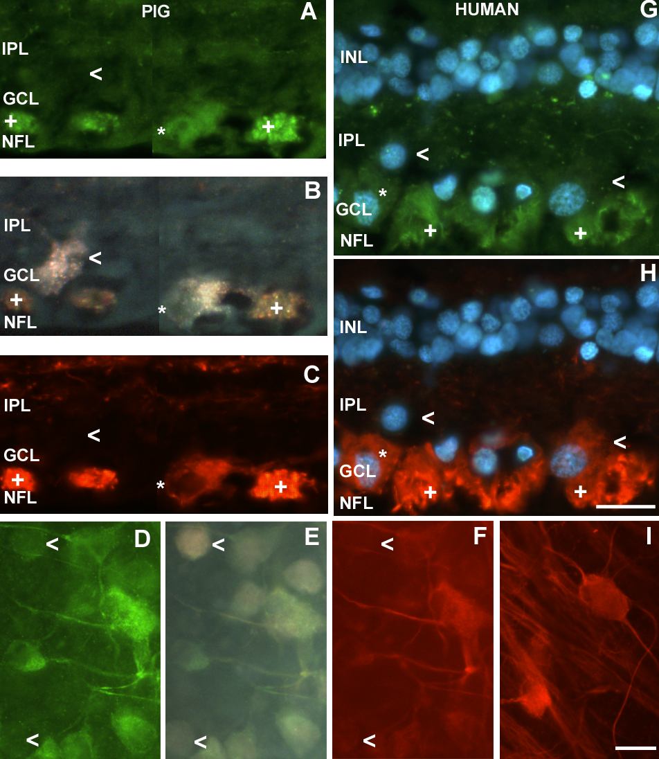
Figure 4. Distribution of NF-L subunit and NF-H
Distribution of NF-L subunit (green) and NF-H (red) in double labeled pig and human retinal sections and flatmounts. A, B and C: Section from pig retina double-immunolabeled with NF-L (A) and NF-H (C), and backfilled with fluorogold (B). The plus symbol (+) indicates immunopositive axon bundles in the nerve fiber layer (NFL), The open arrowhead points to a RGC that was not labelled by either NF-H or NF-L antibodies. An asterisk (*) indicates an RGC in which distribution of NF-L is slightly more internal than that of NF-H. D, E, and F: Flat mount from pig retina double-immunolaled with NF-L (D) and NF-H (F), and backfilled with fluorogold (E). The upper open arrowhead points to a RGC immunoreactive for NF-L but not for NF-H. The lower open arrowhead points to a RGC immunoreactive for NF-H but not for NF-L. G and H: Sections from human retina double-immunolabeled with NF-L (G) and NF-H (H). These antibodies bind axon bundles in the NFL (+), RGC somata and processes in the inner plexiform layer (IPL). The right open arrowhead points to a RGC that stains for NF-H but not NF-L, while the left open arrowhead indicates a RGC that is labeled neither for NF-H nor for NF-L. Nuclei have been labeled with DAPI. An asterisk (*) indicates an RGC in which distribution of NF-L is slightly more internal than that of NF-H. I: Human retina flatmounted, immunostained with NF-H. Homogeneous decoration of RGC somas and dendrites is visible. The scale bar for sections is shown in Panel H and represents 20 μm. The scale bar for flatmounts is shown in Panel I and represents 20 μm. The nerve fiber layer (NFL), ganglion cell layer (GCL), inner plexiform layer (IPL), and inner nuclear layer (INL) are labeled in the images.