![]() Figure 1 of
Ruiz-Ederra, Mol Vis 2004;
10:83-92.
Figure 1 of
Ruiz-Ederra, Mol Vis 2004;
10:83-92.
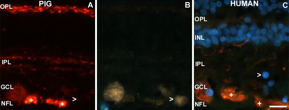
Figure 1. Distribution of NF-H subunit
Distribution of NF-H subunit in pig and human retinal sections. A and B: Sections of pig retina immunostained with NF-H (A) and backfilled with fluorogold (B). NF-H binds the axon bundles in the nerve fiber layer (NFL), retinal ganglion cell (RGC) somata, at least two layers of fibers in the inner plexiform layer (IPL), and the outer plexiform layer (OPL). A few RGC somata are not labeled with NF-H (open arrowhead). C: Section from human retina immunostained with NF-H, nuclei labeled with DAPI. This antibody binds mainly axon bundles in the nerve fiber layer (NFL), RGC somata and some fibers in the IPL. Plus symbols (+) in panels indicate representative axon bundles from the NFL. Open arrowheads point to a RGC soma not labeled with NF-H antibody. The scale bar represents 20 μm. The nerve fiber layer (NFL), ganglion cell layer (GCL), inner plexiform layer (IPL), inner nuclear layer (INL), and outer plexiform layer (OPL) are labeled in the images.