![]() Figure 6 of
Fischer, Mol Vis 2004;
10:973-986.
Figure 6 of
Fischer, Mol Vis 2004;
10:973-986.
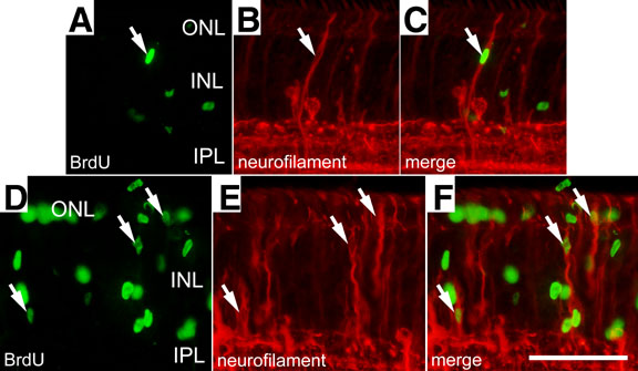
Figure 6. Some of the neurofilament-expressing Müller glia are proliferating
Insulin and FGF2 induce the proliferation of some of the neurofilament expressing Müller glia in peripheral regions of the retina, within 2 mm of the retinal margin. Sections of the retina were labeled with antibodies to BrdU (in green) and neurofilament (NF-M; in red). Eyes received 3 consecutive daily injections of insulin and FGF2 and retinas were obtained at 6 (A-C) or 24 h (D-F) after the final injection. Arrows indicate cells labeled for BrdU and neurofilament. The outer nuclear layer (ONL), inner nuclear layer (INL), and inner plexiform layer (IPL) are labeled. The calibration bar in F represents 50 μm and applies to all panels.