![]() Figure 5 of
Fischer, Mol Vis 2004;
10:973-986.
Figure 5 of
Fischer, Mol Vis 2004;
10:973-986.
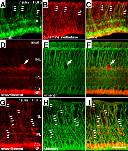
Figure 5. Neurofilament expression co-localizes with glutamine synthetase and vimentin in growth factor treated retinas
Intraocular injections of insulin and FGF2 induce the expression of neurofilament in Müller glia that express glutamine synthetase and vimentin. Retinas were treated with 3 consecutive daily doses of insulin and FGF2 (A-C and G-I) or insulin alone (D-F). Retinas were fixed and processed for immunocytochemistry 24 h after the final dose of growth factors. A-C: Vertical section of the peripheral retina that was labeled with antibodies to neurofilament (NF-M; in green) and glutamine synthetase (in red). D-I: Vertical sections of the retina that were labeled for neurofilament (NF-L; in red) and vimentin (in green). Arrows in A-C and G-I indicate double labeled structures and the arrow in D-F indicates a displaced ganglion cell that is immunoreactive for neurofilament. The inner nuclear layer (INL), inner plexiform layer (IPL), and ganglion cell layer (GCL), and nerve fiber layer (NFL) are labeled. The calibration bar in I represents 50 μm and applies to all panels.