![]() Figure 4 of
Dejneka, Mol Vis 2004;
10:964-972.
Figure 4 of
Dejneka, Mol Vis 2004;
10:964-972.
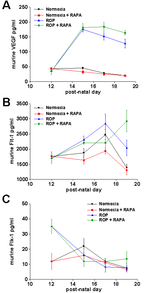
Figure 4. VEGF, Flt-1, and Flk-1 protein levels in developing normal and ROP eye were quantified using ELISA
A: Upon removal of mice from the hyperoxia chamber, VEGF levels increase dramatically. ROP animals continue to maintain high levels of VEGF with rapamycin treatment (4 mg/kg/day). By P19, the level of VEGF in ROP animals receiving drug is higher than that of ROP controls (163.6±7.2 pg/ml compared to 127.8±14.4 pg/ml). B: A general increase in Flt-1 is observed in normoxic and ROP animals from P12-P17, followed by a decrease from P17-P19. High dose rapamycin had no effect on overall levels of Flt-1 in normoxic animals at any time point. In contrast, ROP animals treated with drug exhibit an overall increase in Flt-1 from P17-P19 that is not observed in any other group. C: Hyperoxia causes an increase in Flk-1 in the eye (compare 35.0±4.9 pg/ml [ROP] to 11.8±5.4 pg/ml [normoxia] at P12). By P15 Flk-1 levels in ROP animals drop to those of normoxic controls. Rapamycin (4 mg/kg/day) has no effect on ROP or normoxic animals at any time point examined.
