![]() Figure 3 of
Dejneka, Mol Vis 2004;
10:964-972.
Figure 3 of
Dejneka, Mol Vis 2004;
10:964-972.
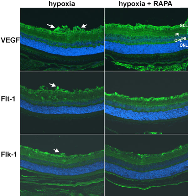
Figure 3. VEGF, Flt-1, and Flk-1 immunostaining in the ROP retina on P19
Magnification of all images are 10x. VEGF, Flt-1, and Flk-1 positive cells are stained in green; Nuclei are labeled blue with DAPI. VEGF protein is localized to cells throughout the eye with the strongest signal in the ganglion cell layer (GCL), surrounding the neovascular tufts (arrows). Rapamycin treatment (2 mg/kg/day or 4 mg/kg/day) has no obvious effect on the overall staining pattern of VEGF in the retina. Flt-1 and Flk-1 are also primarily localized inner plexiform layer (IPL), outer plexiform layer (OPL) and GCL. An increase in Flt-1 is observed with low and high dose rapamycin treatment in the GCL. INL represents inner nuclear layer. Scale bars represent 20 μm.