![]() Figure 1 of
Dejneka, Mol Vis 2004;
10:964-972.
Figure 1 of
Dejneka, Mol Vis 2004;
10:964-972.
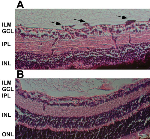
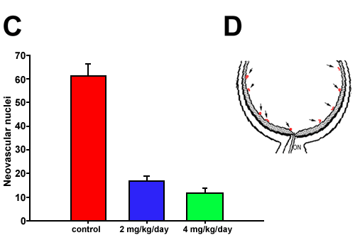
Figure 1. Rapamycin significantly reduces neovascular lesions crossing the ILM in ROP mice
A: Representative section (20x) of a control P19 ROP retina. B: Representative section (20x) from a P19 ROP animal treated with 2 mg/kg rapamycin. Arrows highlight neovascular tufts protruding into the vitreous. Scale bars in A and B represent 40 μm. C: The average number of vascular nuclei found anterior to the ILM per 5 μm cross section were determined for each experimental group. The error bars represent (SEM). D: Cartoon indicating location of pathologic neovascular nuclei. Arrow, neovascular fronds; ON, optic nerve.