![]() Figure 9 of
Stark, Mol Vis 2004;
10:943-955.
Figure 9 of
Stark, Mol Vis 2004;
10:943-955.
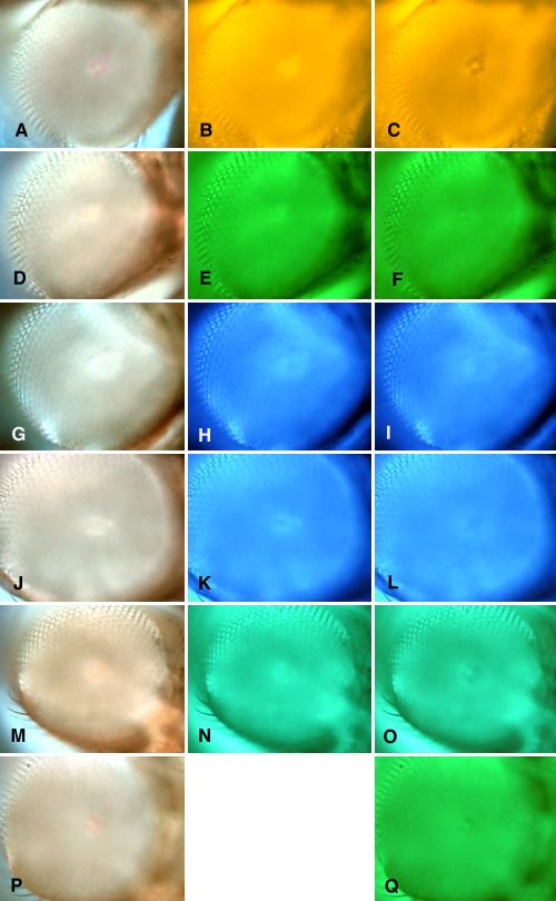
Figure 9. Imaging of visual pigment and rhodopsin-metarhodopsin conversions
The deep pseudopupil was used to image visual pigment and rhodopsin-metarhodopsin conversions in normal flies (w, row 1) and transgenic flies with Rh2 to Rh6 replacing Rh1 in R1-6 (rows 2 to 6), respectively. Native Rh1 in R1-6 viewed with white light (A), 579 nm light (B), and 579 nm light darkened with 460 nm light (C). Transgenic with Rh2 in R1-6, viewed with white light (D), 520 nm light (E), and 520 nm light darkened with 436 nm light (F). Transgenic with Rh3 viewed with white light (G), 480 nm light (H), and 480 nm light darkened with 350 nm light. Transgenic with Rh4 viewed with white light (J), 480 nm light (K), and 480 nm light darkened with 376 nm light (L). Transgenic with Rh5 viewed with white light (M), 505 nm light (N), and 505 nm light darkened with 405 nm (O). Transgenic with Rh6 viewed with white light (P), and with 524 nm light (Q).