Figure 11 of Stark, Mol Vis 2004; 10:943-955.
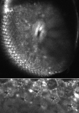
Figure 11. R1-6 GFP (confocal microscopy)
Deep pseudopupil (top) and optical neutralization of the cornea (bottom) of typical white eyed fly from the cross that gives GFP expression in R1-6.