![]() Figure 8 of
Khanobdee, Mol Vis 2004;
10:933-942.
Figure 8 of
Khanobdee, Mol Vis 2004;
10:933-942.
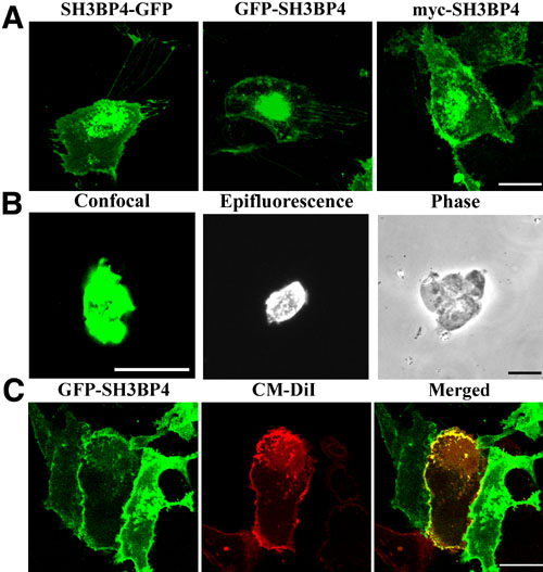
Figure 8. Confocal photomicrographs of APRE-19 transfected with SH3BP4-GFP, GFP-SH3BP4, and myc-SH3BP4 constructs
A: Plasma membrane and nuclear periphery localization of the SH3BP4 fusion proteins. B: Nuclei from ARPE-19 transfected with GFP-SH3BP4 for 24 h were purified using NE-PER reagent. Confocal and epifluorescence microscopy show GFP-SH3BP4 at the nuclear periphery. The corresponding phase microscopy image for the epifluorescent nuclei is also shown. C: Confocal photomicrographs of APRE-19 transfected with GFP-SH3BP4 for 24 h and stained with the plasma membrane marker CM-DiI. GFP-SH3BP4 at the cell periphery co-localizes with the plasma membrane marker CM-DiI seen in yellow in the merged image. Bars represent 20 μm.