![]() Figure 7 of
Khanobdee, Mol Vis 2004;
10:933-942.
Figure 7 of
Khanobdee, Mol Vis 2004;
10:933-942.
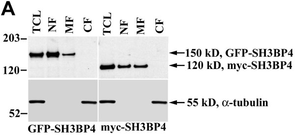
Figure 7. Subcellular fractionation of SH3BP4 fusion proteins
A: ARPE-19 cells were transfected with either GFP-SH3BP4 or myc-SH3BP4 constructs for 24 h followed by differential centrifugation subcellular fractionation. Fractions were analyzed by immunoblot for SH3BP4. B: Qualitative analysis of these data were conducted for SH3BP4 expression relative to μg of total protein. The fold level increases between the fractions are shown above each bar; levels that were not detectable (ND) are also noted.