![]() Figure 4 of
Khanobdee, Mol Vis 2004;
10:933-942.
Figure 4 of
Khanobdee, Mol Vis 2004;
10:933-942.
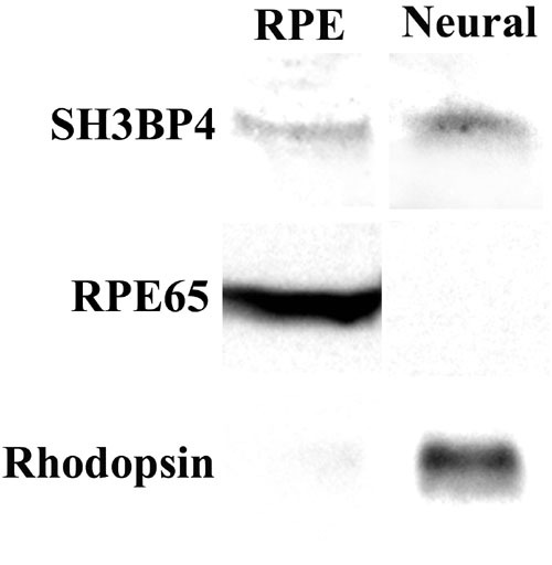
Figure 4. Western blot analysis of rabbit retinas
Six rabbit eyes were dissected into RPE and neural retina layers. Tissue lysates were immunoblotted using a 1:5000 dilution of SH3BP4, a 1:5000 dilution of RPE65 or a 1:2000 dilution of rhodopsin antibodies. SH3BP4 was detected in all 12 samples and one representative lane is shown. Each lane contains 30-33 μg of total protein. Immunoblots are shown for SH3BP4, for the RPE control protein (RPE65) and the neural retina control protein, rhodopsin. The rhodopsin monomer band at about 35 kDa is shown in the figure but monomers, dimers, and tetramers of rhodopsin were detected on the immunoblot.