![]() Figure 2 of
Khanobdee, Mol Vis 2004;
10:933-942.
Figure 2 of
Khanobdee, Mol Vis 2004;
10:933-942.
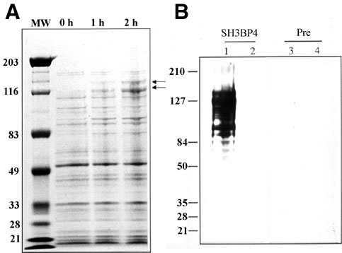
Figure 2. Expression and detection of the MAL-SH3BP4 fusion protein
A: SH3BP4 was cloned into the pMAL vector and expressed in XL10-Gold bacterial cells. Cells were grown to an A600 of about 0.5 followed by treatment with IPTG for 0, 1, or 2 h. Cells were lysed in 2% SDS with protease inhibitors, run on a 4-12% Bis-Tris gel and stained with Coomassie Blue. The double arrows show the Coomassie blue stained MBP-SH3BP4 fusion protein that is visible in the 1 h and 2 h induced bacterial lysates and also point to the region that was excised (from a separate gel) and used as the antigen for raising the SH3BP4 antibody. B: Cells were induced with IPTG for 0 h (lanes 2, 4) or for 2 h (lanes 1, 3), lysed, run on a 4-12% Bis-Tris gel, transferred to nitrocellulose and stained with a 1:50,000 dilution of the chicken anti-human SH3BP4 IgY (lanes 1, 2) or a 1:10,000 dilution of the pre-immune IgY (lanes 3, 4).