![]() Figure 8 of
Tanemura, Mol Vis 2004;
10:923-932.
Figure 8 of
Tanemura, Mol Vis 2004;
10:923-932.
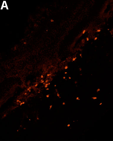
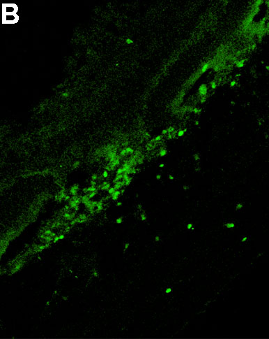
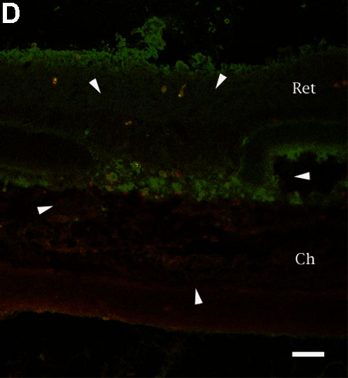
Figure 8. Detection and localization by immunofluorescent labeling of VEGFR2 and ERβ by a confocal laser scanning microscope
Frozen section of a female rat eye 3 days after laser photocoagulations. Lesions surrounded by arrowheads show the CNV lesion. The choroid (Ch) and retina (Ret) are labeled around the lesions. A: VEGFR2 was labeled using anti-VEGFR2 monoclonal IgG, and TRITC conjugated rabbit anti-mouse IgG. B: ERβ was labeled using anti-ERβ rabbit IgG and FITC conjugated swine anti-rabbit IgG. C: Overlaid image of A and B. Co-labeled cells are marked with arrows in the overlaid image. Scale bar represents 50 μm. D: No staining was observed in the control section. Scale bar represents 50 μm.