![]() Figure 1 of
Tanemura, Mol Vis 2004;
10:923-932.
Figure 1 of
Tanemura, Mol Vis 2004;
10:923-932.
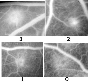
Figure 1. Typical examples of each CNV score in the fluorescein angiogram after photocoagulation in rat retinae
Each photocoagulated spot was scored from 0 to 3 according to the size and the presence or absence of dye leakage on the early and late phase angiogram photographs. The guideline for the CNV scoring was as follows: no leakage (score 0); minimum leakage or a staining of tissue with no leakage (score 1); small but evident leakage less than 1/4 disc area (indicating small active CNV; score 2); large evident leakage (large active CNV; score 3). Overall photo-intensity was normalized by retinal capillary fluoro-intensity.