![]() Figure 7 of
De Maria, Mol Vis 2004;
10:74-82.
Figure 7 of
De Maria, Mol Vis 2004;
10:74-82.
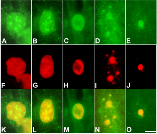
Figure 7. DNase I distribution during nuclear breakdown
A-E: DNase I immunolabeled with antibody Ab2983. F-J: Hoechst 33342 (showed in red). K-O: Merge of Ab2983 and Hoechst 33342 labeling. A,F,K: Nucleus from a young fiber located at the outer cortex. B,G,L: Nucleus from a fiber located at the mid cortex. C-D,H-I,M-N: Nuclear material from fibers located at the inner cortex. E,J,O: Nuclear remnants from a fiber located at the deep inner cortex. The nuclear localization of DNase I changes with the progression of nuclear breakdown. The enzyme is closely associated with highly condensed chromatin at the late stages of the process. Scale bar represents 5 μm.