![]() Figure 6 of
De Maria, Mol Vis 2004;
10:74-82.
Figure 6 of
De Maria, Mol Vis 2004;
10:74-82.
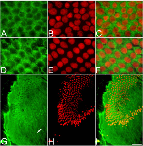
Figure 6. DNase I distribution during fibergenesis
A-F: Whole mounts of lens capsule with adhered fibers. G-I: Superficial lens cryosections. A,D: DNase I immunolabeled with antibody Ab2983 and G with antibody AbR. B,E,H: Hoechst 33342 (showed in red). C,F,I: Merge of a-DNase I antibodies and Hoechst 33342 labeling. Note that a fraction of DNase I is located at the nuclear territory in elongating fibers. Scale bar represents 10 μm in A-F and 60 μm in G-I.