![]() Figure 3 of
De Maria, Mol Vis 2004;
10:74-82.
Figure 3 of
De Maria, Mol Vis 2004;
10:74-82.
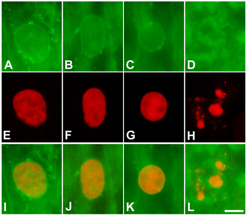
Figure 3. Nuclear membrane in cortical fibers
A-D: Membrane labeling with DiOC6. E-H: Hoechst 33342 (red). I-L: Merge of DiOC6 and Hoechst 33342. A,E,I: Nucleus from a fiber located at the outer cortex. B,F,J: Nucleus from a fiber located at the mid cortex. C,G,K,D,H,L: Nuclear material from fibers located at the inner cortex. Nuclear membrane degradation begins after the onset of chromatin condensation and DNA cleavage. Scale bar represents 4 μm.