![]() Figure 2 of
De Maria, Mol Vis 2004;
10:74-82.
Figure 2 of
De Maria, Mol Vis 2004;
10:74-82.
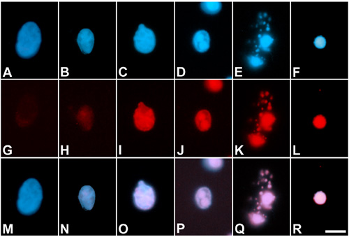
Figure 2. Chromatin condensation and fragmentation during nuclear breakdown
A-F: Hoechst 33342 staining. G-L: TUNEL assay. M-R: Merge of Hoechst 33342 and TUNEL. A,G,M: Nucleus from a fiber located at the mid cortex. B,H,N: Nucleus from a deeper fiber. C-E,I-K,O-Q: Nuclear material in fibers located at the inner cortex. F,L, and R: Nuclear remnant from a fiber located at the deep inner cortex. During the process of nuclear breakdown chromatin condensation and fragmentation increase and different patterns of condensation can be observed. Scale bar represents 5 μm.