![]() Figure 1 of
De Maria, Mol Vis 2004;
10:74-82.
Figure 1 of
De Maria, Mol Vis 2004;
10:74-82.
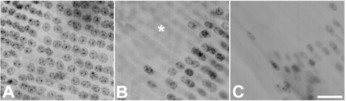
Figure 1. Nuclear shape during early stages of fibergenesis
Whole mounts of lens capsules with adhered cells stained with Ferric hematoxylin. A: Round shaped nuclei in post mitotic cells (upper left) and in earliest fibers at meridional rows. B: Round shaped nuclei in the outermost cortical fibers. C: Elongated nuclei from fibers located at the outer cortex. Note that nuclear shape changes from round to elongate with progression of fiber differentiation. The asterisk marks a region of aligned post-mitotic epithelial cells seen through the fibers. Scale bar represents 20 μm in A,B and 30 μm in C.