![]() Figure 4 of
Chiambaretta, Mol Vis 2004;
10:901-909.
Figure 4 of
Chiambaretta, Mol Vis 2004;
10:901-909.
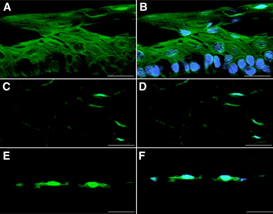
Figure 4. Cell and tissue immunolocalization of KLF4 proteins in human ocular cornea
KLF4 immunolocalization (green fluorescence staining) was performed in human corneal epithelium (A,B), corneal stroma (C,D), and corneal endothelium (E,F). Cell nuclei were visualized with DAPI (blue staining). A,C,E: Green only images related to KLF4 proteins. B,D,F: Green-blue images related to mix of KLF4 and DAPI staining. Acquisitions were made under standard conditions with a fluorescence Axiophot microscope (Zeiss). Magnification is x400. Scale bar represents 10 μm.