![]() Figure 3 of
Chiambaretta, Mol Vis 2004;
10:901-909.
Figure 3 of
Chiambaretta, Mol Vis 2004;
10:901-909.
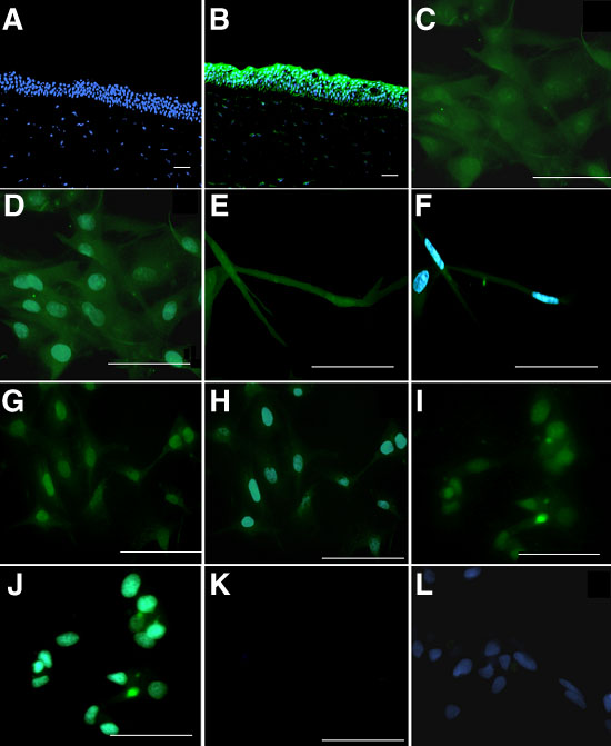
Figure 3. Cell and tissue immunolocalization of KLF4 proteins in human ocular surface
KLF4 immunolocalization (green fluorescence staining) was performed in human cornea (B), HCE cell line (C,D), human corneal keratocytes (E,F), HTEC cell line (G,H), and human conjunctival epithelial cell line (I,J). Negative controls (A,K) were obtained with the same conditions but in the absence of KLF4 antibody incubations. Cell nuclei were visualized with DAPI (blue staining; A,B,D,F,H,J,L). Acquisitions were made under standard conditions with a fluorescence Axiophot microscope (Zeiss). Magnifications for A and B are x40, and for C-L are x200. Scale bar represents 10 μm.