![]() Figure 2 of
Zhang, Mol Vis 2004;
10:884-889.
Figure 2 of
Zhang, Mol Vis 2004;
10:884-889.
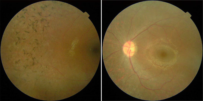
Figure 2. Fundus photographs
Fundus photographs from individual 11 showing typical changes of RP including a waxy pale optic disc, attenuation of retinal arteries, and bone spicule pigment deposits in the mid-periphery of the retina. Left: Right eye showing macula and mid-periphery retina. Right: Left eye showing optic disc, retinal vessels and macular.