![]() Figure 6 of
Chen, Mol Vis 2004;
10:874-883.
Figure 6 of
Chen, Mol Vis 2004;
10:874-883.
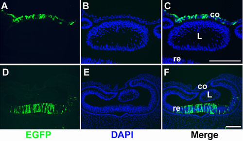
Figure 6. Transfection of corneal epithelial and retinal neuroepithelial cells with EGFP
Chick embryos at stage 15 were used for electroporation. For the corneal epithelial cells (A-C), pCAX DNA was placed by the surface ectoderm at the eye region. For retinal neuroepithelial cell electroporation (D-F), DNA was injected into the vitreous cavity. After incubation for 24 h, embryos at stage 21 were collected for EGFP expression analysis (shown in A and D). Cell nuclei were stained with DAPI (B and E). Merged images of A,B and D,E are shown in C and F, respectively. Scale bars represent 100 μm.