![]() Figure 4 of
Chen, Mol Vis 2004;
10:874-883.
Figure 4 of
Chen, Mol Vis 2004;
10:874-883.
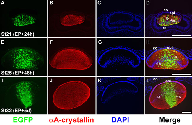
Figure 4. EGFP expression in lens fiber cells
pCAX DNA was electroporated into posterior lens vesicle cells using chicken embryos at stage 15. After incubation for 24 h (A-D), 48 h (E-H), or 5 days (I-L), embryos were harvested and stained for EGFP (A, E, I) or αA-crystallin (E, F, J). Cell nuclei were revealed by DAPI staining (C, G, and K). Merged images are shown in D, H, and L. A-D: At stage 21 (st21), about 24 h post-electroporation (EP+24h), a high percentage of lens fiber cells expressed EGFP. E-H: At stage 25 (st25), 48 h post-electroporation (EP+48h), the EGFP expressing fiber cells were localized toward the center of the lens, due to the maturation process of lens fiber cells. I-L: At stage 32 (st32), 5 days after electroporation (EP+5d), the transfected lens fiber cells, located at the center of the lens, still express high levels of EGFP. Scale bars represent 100 μm.