![]() Figure 3 of
Chen, Mol Vis 2004;
10:874-883.
Figure 3 of
Chen, Mol Vis 2004;
10:874-883.
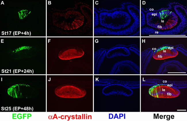
Figure 3. Targeted expression of EGFP in lens epithelial cells
pCAX DNA was introduced into the anterior lens cells by electroporation using chicken embryos at stage 15. Injected embryos were returned to incubation and then harvested at 4 h (A-D), 24 h (E-H), or 48 h (I-L) after electroporation at stage 17, 21, and 25, respectively. Embryos were briefly fixed with 10% formalin and 10 μm frozen sections were cut to stain for EGFP (green in A, E, I), αA-crystallin (red in B, F, J) and nuclei (blue DAPI staining in C, G, K). Merged images are shown in D, H and L. A-D: At stage 17 (st17) 4 h post-electroporation (EP+4h), EGFP expression can be detected in some of the anterior cells of the lens vesicle. At this stage, αA-crystallin is weakly expressed in the posterior lens vesicle cells. E-H: At stage 21 (st21), about 24 h post-electroporation (EP+24h), the posterior lens cells have elongated and differentiated into the fiber cells which express high levels of αA-crystallin (F). EGFP positive cells were localized in the anterior lens epithelial cells. I-L: At stage 25 (st25), 48 h post-electroporation (EP+48h), EGFP expression was seen in lens epithelial cells and in fiber cells at the equatorial region, suggesting that some of the EGFP positive epithelial cells had differentiated into fiber cells. The cornea (co), lens (le), lens epithial cells (epi), lens fiber cells (fib), retina (re) are labeled in this figure. Scale bars represent 100 μm.