![]() Figure 2 of
Chen, Mol Vis 2004;
10:874-883.
Figure 2 of
Chen, Mol Vis 2004;
10:874-883.
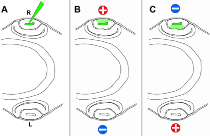
Figure 2. Schematic illustration of microinjection and electroporation to target EGFP expression to the lens
A: A glass needle filled with plasmid DNA mixed with fast green dye is inserted into the lens vesicle of the right eye which faces up toward the eggshell. After DNA is injected into the lumen of the lens vesicle, the glass needle is retracted before electroporation. B: To electroporate the lens epithelial cells, the anode (+) is placed above the right eye and the cathode (-) underneath the left eye. The electric current drives DNA, which is negatively charged, into the lens epithelial cells (represented by the light green area). C: To introduce DNA into the lens fiber cells, the placement of the electrodes is reversed.