![]() Figure 3 of
Boehlke, Mol Vis 2004;
10:867-873.
Figure 3 of
Boehlke, Mol Vis 2004;
10:867-873.
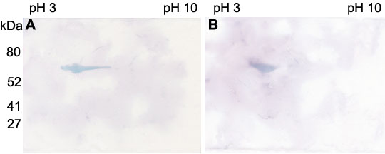
Figure 3. Western blot analysis of corneal epithelial cell lysates subjected to two dimensional gel electrophoresis
Isoelectric focusing by pH was run horizontally and the SDS-PAGE by molecular weight was run vertically. Acidic proteins are on the left and basic proteins are on the right. A: E-17 antibody recognized a protein band in the acidic region at about 55 kDa. B: J7 antibody also reacted with an acidic protein with a molecular weight of about 55 kDa.