![]() Figure 2 of
Boehlke, Mol Vis 2004;
10:867-873.
Figure 2 of
Boehlke, Mol Vis 2004;
10:867-873.
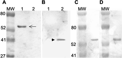
Figure 2. Western blot analyses of cell lysates by E17 antibody and J7 (anti-cytokeratin 12) antibody
A: Western blot analysis showed that E-17 antibody recognized a protein of 55 kDa (arrow) in corneal epithelial lysates(lane 1), but did not identify such a protein in lysates of HEK 293 cells transfected with Gαq cDNA (lane 2). B: Western blot analysis showed G4415 anti-Gαq antibody recognized a protein of 42 kDa only in lysates of HEK 293 transfected with Gαq cDNA (arrowhead, lane 2), but not in corneal epithelial cells (lane 1). C: Western blot analysis of corneal epithelial lysates stained with E-17 antibody. D: Western blot analysis of corneal epithelial lysates stained with J7 (anti-CK12) antibody. Both antibodies (E-17 in C and J7 in D) identified equivalent 55 kDa bands and reconfirmed the finding in A that E-17 reacted with the 55 kDa protein (cytokeratin 12), instead of the purported Gαq (42 kDa).