![]() Figure 1 of
Boehlke, Mol Vis 2004;
10:867-873.
Figure 1 of
Boehlke, Mol Vis 2004;
10:867-873.
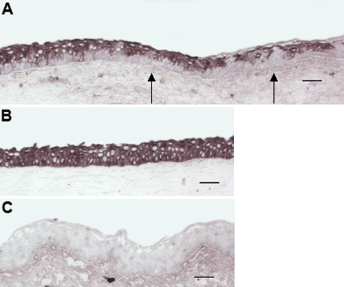
Figure 1. Immunohistochemical analysis of human ocular surface epithelia with anti-Gαq antibody (E-17)
Immunohistochemistry of human ocular surface epithelia with anti-Gαq antibody (E-17) showed a preferential staining of the suprabasal limbal epithelium (A) and the full layer of central corneal epithelium (B), with complete absence of staining in the conjunctival epithelium (C) and basal limbal epithelium (A, arrows). Scale bars represent 50 μm.