![]() Figure 3 of
Feng, Mol Vis 2004;
10:845-850.
Figure 3 of
Feng, Mol Vis 2004;
10:845-850.
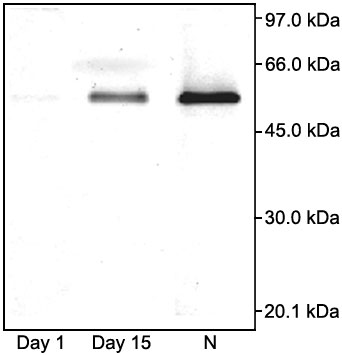
Figure 3. ALDH3A1 enzyme activity is restored after cornea healing
ALDH3A1 specific substrate zymography of 10 μg water soluble protein from mouse cornea of normal control (N), at Day 1, and Day 15 following alkali burn. The size and position of markers are shown on the right. Protein samples were separated by 10% native SDS-PAGE and after rinsing out SDS from the gel the ALDH3A1 band was developed for 15 min at 37 °C with 10 ml of 10 mM phosphate buffer (pH 7.0) solution containing 20 mg NADP+, 8 mg MTT, 0.4 mg phenazine methosulfate and 20 μl benzaldehyde.