![]() Figure 5 of
Wong, Mol Vis 2004;
10:837-844.
Figure 5 of
Wong, Mol Vis 2004;
10:837-844.
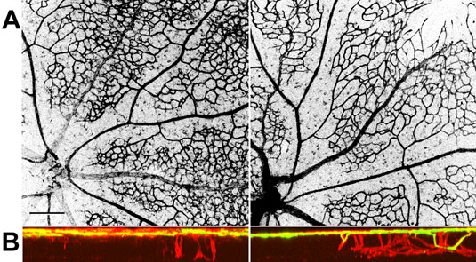
Figure 5. PEDF over-expression slows the rate of vessel differentiation and growth of vessels into the deeper layers of the retina
A: Extended focus confocal images of the structure of vascular trees in whole mount retinae from a P12 animal injected subretinally at P2. Vascular structures are visualized by immunohistochemistry with isolectin GS-IB4, which specifically labels endothelial cells. The retina injected with AAV2/1.CMV.PEDF (experimental eye) is shown on the left, and the fellow eye injected with AAV2/1.CMV.EGFP (control eye) is shown on the right. In both retinae, there is differentiation of vessels into arterioles (thinner vessels) and venules (thicker vessels). However, in the experimental eyes (left), the presumptive capillary structure retains a denser, more immature, polygonal vascular pattern compared to the control eyes (right). In the control eyes, there is a slightly sparser, more differentiated branching structure. B: The confocal cross-section reconstructions in the same retinae corresponding to those shown in the above in A. The yellow signal shows the distribution of fluorescein dextran after intracardiac injection, indicating the distribution of patent vessels in the vascular tree. The red signal shows the pattern of GS-IB4 labeling, indicating the structure of both patent vessels as well as nascent vessels that have not yet undergone cannulation. The experimental eyes (left) show a sparser distribution of nascent vessels in the deeper layers of the retina; many of these vessels have not yet undergone cannulation. However, in the control contralateral eyes, there is a greater number of vessels growing into the deeper layers with some vessels having already undergone cannulation.