![]() Figure 1 of
Wong, Mol Vis 2004;
10:837-844.
Figure 1 of
Wong, Mol Vis 2004;
10:837-844.
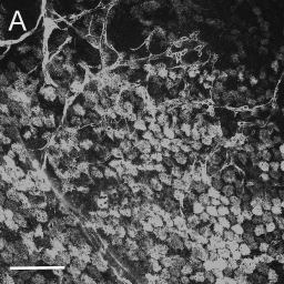
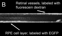
Figure 1. Robust expression of viral transgenes following subretinal injection AAV.
Confocal micrograph showing high levels of EGFP in the RPE cell layer of a P9 mouse that was injected subretinally with AAV2/1.CMV.EGFP at P2. A: An extended focus (merged z-stack) confocal image of this whole mount retina. The tissue was illuminated with the 488 nm line of the krypton-argon laser and the resulting emission viewed through a 522/35 nm filter. Retinal vessels are rendered visible by the perfusion of fluorescein dextran. Green fluorescence was clearly visualized in the cytoplasm of cells in the RPE. Scale bar represents 100 μm. B: The same z-stack of confocal images now viewed in cross-section. The fluorescent signal deep to the retina (indicated by the lower arrow) arises from the RPE layer. This layer is distinct from the fluorescence from the fluorescein labeled retinal vessels.