![]() Figure 6 of
Brossas, Mol Vis 2004;
10:65-73.
Figure 6 of
Brossas, Mol Vis 2004;
10:65-73.
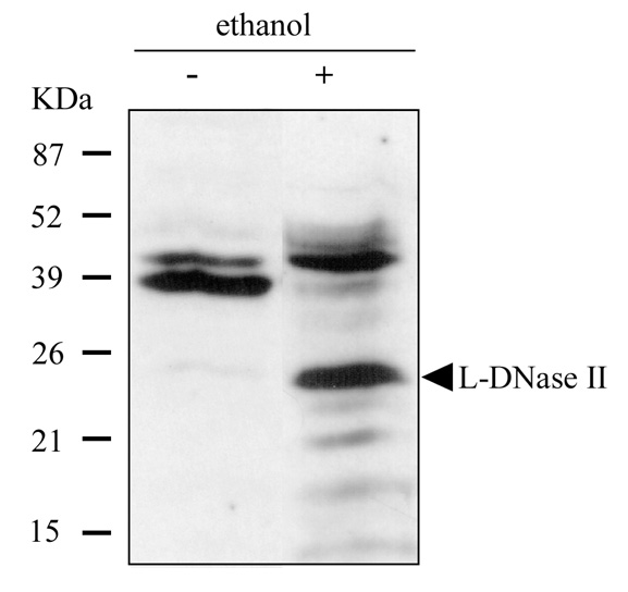
Figure 6. Western blot analysis of L-DNase II expression in ARPE-19 cells
The total protein extracts were separated on 13% SDS-PAGE gel, transferred, and stained by enhanced chemiluminescence method with a polyclonal antibody directed against 181-191 peptide of LEI. The extracts were isolated from cells that had been incubated without (lane 1) or with 10% ethanol (lane 2) for 24 h. Each lane of the gel was loaded with 40 microgram of protein. The molecular weight is indicated on the left.