![]() Figure 5 of
Brossas, Mol Vis 2004;
10:65-73.
Figure 5 of
Brossas, Mol Vis 2004;
10:65-73.
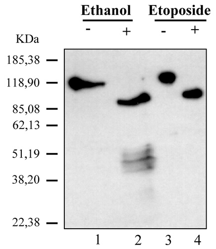
Figure 5. Western blot analysis of PARP cleavage in ARPE-19 cells
Nuclear protein extract was separated on 8% SDS-PAGE gel, transferred, and stained by enhanced chemiluminescence method with a polyclonal anti-PARP C-2-10 antibody. Nuclear proteins were isolated from cells that had been incubated without (lane 1) or with 10% ethanol for 24 h (lane 2). An untreated HL-60 cell lysate (lane 3) and an HL-60 cell lysate treated with etoposide (provided by Zymed Laboratories) to induce cell death (lane 4), represented un-cleaved and cleaved PARP respectively. Lanes 1 and 2 of the gel were loaded with 5 microgram protein. Molecular weight markers are indicated on the left.