![]() Figure 2 of
Brossas, Mol Vis 2004;
10:65-73.
Figure 2 of
Brossas, Mol Vis 2004;
10:65-73.
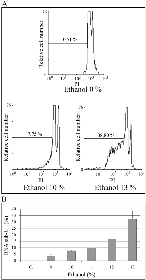
Figure 2. DNA degradation in ARPE-19 cells induced to die by ethanol
Panel A: Typical pattern of DNA fluorescence histograms (log scale) of propidium iodide-stained ARPE-19 cells grown in presence of 0% (top), 10% (left) and 13% (right) ethanol. The area located to the left of the G1 peak is used to calculate DNA degradation. Panel B: DNA degradation histograms of ARPE-19 subject to different concentrations of ethanol. DNA sub G1 experimental values are means±standard deviation of three different experiments performed for each ethanol concentration.