![]() Figure 1 of
Brossas, Mol Vis 2004;
10:65-73.
Figure 1 of
Brossas, Mol Vis 2004;
10:65-73.
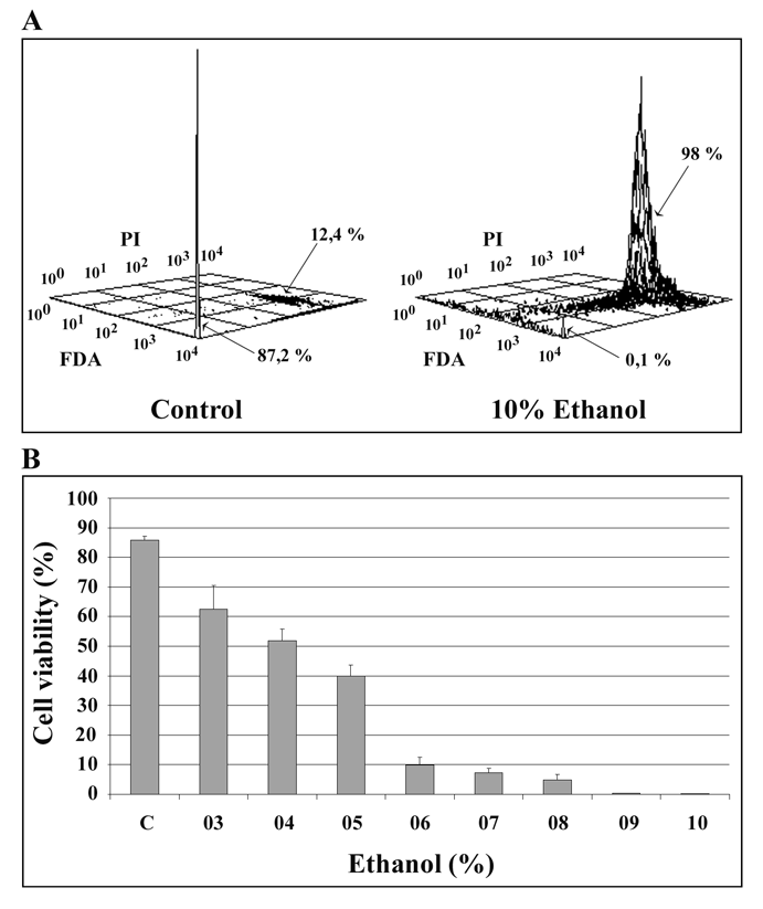
Figure 1. Effects of ethanol on ARPE-19 cell viability
Cells were treated with different concentrations of ethanol, stained with FDA and PI, and quantified by flow cytometry analysis. Panel A: The x-axis represents green fluorescence of FDA, the y-axis represents red fluorescence of PI, and the z-axis represents cell number in arbitrary units. Panel B:The percentage of viable cells was calculated and reported as a histogram. Values are means±standard deviation of three different experiments performed for each ethanol concentration. Each histogram was generated with at least 10,000 cells.