![]() Figure 7 of
Cornish, Mol Vis 2004;
10:1-14.
Figure 7 of
Cornish, Mol Vis 2004;
10:1-14.
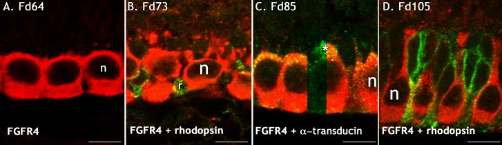
Figure 7. Cones, but not rods, are FGFR4 immunoreactive in fetal monkey retina
FGFR4-IR (red) in sections double labelled with anti-rhodopsin (green; B, D) and antibody to the cone-specific marker, α-transducin (green; C). A: Cones in the incipient fovea are strongly immunoreactive to FGFR4 at Fd 64, when very few cones are immunoreactive to L/M opsin. B: A slightly oblique section on the edge of the incipient fovea shows a small number of rhodopsin-IR cells in the ONL which did not colocalize with FGFR4. C: FGFR4-IR cells colocalized with the cone marker, α-transducin. D: FGFR4-IR cells had a distinctive cone morphology and were interspersed with rhodopsin-IR rods on the edge of the incipient fovea. No cells colocalized rhodopsin and FGFR4. All scale bars represent 10 μm. The cone nuclei (n) and a rod (r) are also identified.