![]() Figure 6 of
Cornish, Mol Vis 2004;
10:1-14.
Figure 6 of
Cornish, Mol Vis 2004;
10:1-14.
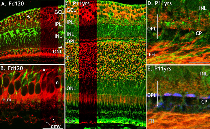
Figure 6. FGFR4 immunoreactivity in monkey fetal and postnatal central retina
FGFR4 immunoreacitivity (red) in fetal and adult macaque retina, immunolabelled with anti-vimentin (green; A-E) and anti-synaptophsyin (blue; E). A: FGFR4-IR in ganglion cell somata, processes and in cones on the edge of the developing fovea. B: A high magnification of the outer retina at the developing fovea showing FGFR4-IR cone somata and pedicles (asterisk). C: FGFR4-IR on the rim of adult fovea is intense in the cytoplasm of ganglion cells and in presumed ganglion cell dendrites in the IPL. FGFR4-IR is present in the OPL, along the length of fibers of Henle and in the inner segments of photoreceptors. The green labelling in the RPE and at the level of the IS and OS is non-specific. D: A high power view in adult retina shows FGFR4-IR in the fibers of Henle, and mild IR associated with the bases of cone pedicles and in the OPL. E: Synaptophysin-IR colocalizes with FGFR4 on the bases of the pedicles. All scale bars represent 20 μm. The cone pedicle (CP), fibers of Henle (FH), ganglion cell layer (GCL), inner nuclear layer (INL), inner plexiform layer (IPL), outer plexiform layer (OPL), presumed developing microvilli (dmv), and external limiting membrane (elm) are also identified.