![]() Figure 5 of
Cornish, Mol Vis 2004;
10:1-14.
Figure 5 of
Cornish, Mol Vis 2004;
10:1-14.
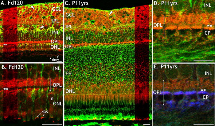
Figure 5. FGFR3 immunoreactivity in monkey fetal and postnatal central retina
FGFR3-IR (red) in fetal and adult macaque retina, immunolabelled with anti-vimentin (green; A-E) and anti-synaptophsyin (blue; E). A: FGFR3-IR on the foveal rim. Müller cell somata and processes colocalize vimentin and FGFR3 (oblique arrowheads). The OPL is almost completely labelled except for a narrow band adjacent to the cone pedicles (double asteriks). B: A high magnification showing FGFR3-IR in the INL, OPL, and ONL, and indicating (double asterisks) the non-reactive band. Developing outer segments of some cones are also FGFR3-IR. C: FGFR3-IR is intense in the majority of cells in the GCL, in isolated cells in the INL, in the majority of ONL somata, in the inner segments of photoreceptors as well as in the IPL and OPL on the foveal rim. The proximal parts of FH are also immunoreactive. The green labelling in the RPE and at the level of the IS and OS is non-specific. D: A high power view showing FGFR3-IR in the OPL and on the bases of the cone pedicles. A non-reactive band, about 2 μm deep, is indicated (double asterisks) E: The same section after labelling with showing co-localization of FGFR3 with synaptophysin on the bases of the pedicles. All scale bars represent 20 μm. The cone pedicle (CP), developing outer segments (dOS), fibers of Henle (FH), ganglion cell layer (GCL), inner nuclear layer (INL), inner plexiform layer (IPL), and nuclei (n) are also identified.