![]() Figure 4 of
Cornish, Mol Vis 2004;
10:1-14.
Figure 4 of
Cornish, Mol Vis 2004;
10:1-14.
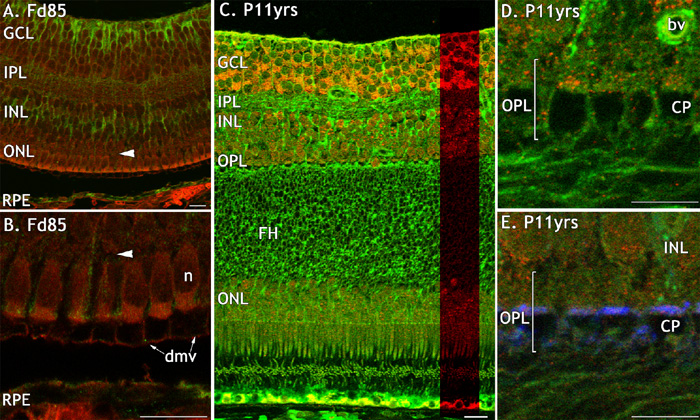
Figure 4. FGFR2 immunoreactivity in monkey fetal and postnatal central retina
FGFR2-IR (red) in fetal and adult macaque retina, double labelled with anti-CRALBP (green; A, B), anti-vimentin (green; C-E) and anti-synaptophsyin (blue; E). A: Low FGFR2 immunoreactivity in all retinal layers and in the RPE at the incipient fovea. Horizontal arrowhead shows the position of the developing OPL (A,B). B: A high magnification of the outer retina and RPE. C: FGFR2-IR is intense in the GCL on the rim of adult fovea, and at lower levels in the INL and ONL. The green labelling in the RPE and at the level of the IS and OS is non-specific. D: A high power view of the OPL in adult retina showing weak FGFR2-IR in the OPL. E: There is a virtual absence of FGFR2 on the cone pedicles and axons. All scale bars represent 20 μm. The cone pedicle (CP), ganglion cell layer (GCL), inner nuclear layer (INL), inner plexiform layer (IPL), inner segment (IS), presumed developing microvilli (dmv), nerve fiber layer (NFL), retinal pigmented epithelium (RPE), blood vessel (bv), and cone nuclei (n) are also identified.