![]() Figure 2 of
Cornish, Mol Vis 2004;
10:1-14.
Figure 2 of
Cornish, Mol Vis 2004;
10:1-14.
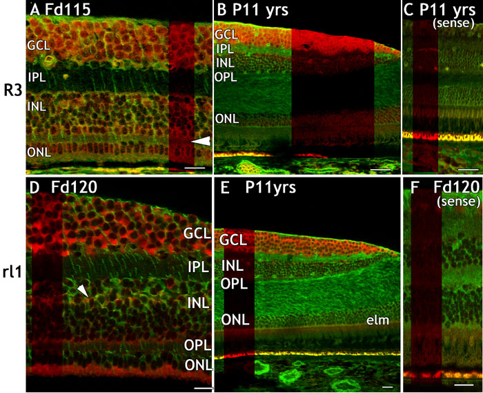
Figure 2. Distribution of FGFR3 and FGFrl1 mRNAs in fetal and postnatal monkey retina
Sections of fetal and adult monkey retina hybridized using human RNA probes to show expression of FGFR3 (A-C) or FGFrl1 (D-F) mRNA (red) and vimentin-IR (green). A: Cytoplasmic and nuclear FGFR3 mRNA is abundant in virtually all cells in the developing retina. Expression in Müller cell somata could not be ruled out, but FGFR3 mRNA was not detected in Müller cell processes. Horizontal arrowhead indicates the position of the developing OPL. B: FGFR3 mRNA is predominantly in ganglion cells in the adult. C: FGFR3 sense probe. The apparent labelling of the RPE is autofluorescence; some capillaries bound the sense probe but neural elements were not labelled. D: FGFrl1 mRNA is expressed in ganglion cells in the developing fovea, in some INL cells, including Müller cells that co-localize vimentin (oblique arrowhead), and the inner segments of photoreceptors, including cones. E: FGFrl1 expression is virtually confined to the GCL in the adult. F: FGFrl1 sense probe. Apparent labelling in the RPE is autofluorescence. All scale bars represent 20 μm. The ganglion cell layer (GCL), inner nuclear layer (INL), inner plexiform layer (IPL), outer nuclear layer (ONL), outer plexiform layer (OPL), and external limiting membrane (elm) are also identified.