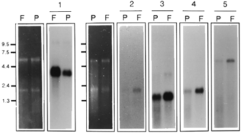
Five micrograms of total RNA per lane from pooled human fovea (F) or pooled human midperipheral retina (P) were submitted to northern analysis. Photographs of the preparative agarose gel used for northern blot analysis showing ethidium bromide stained total RNA from fovea and midperipheral retina is shown to the left hand side of the corresponding panels of blot analysis. Clones representing new human ESTs are indicated (*).
Panel 1) Autoradiograph of blot probed with HFD122210 (Calmodulin, 2 day exposure).
Panel 2) Autoradiograph of blot probed with HFD121216 (NADH 4, 20 min exposure).
Panel 3) Autoradiograph of blot probed with HFD122215* (2 day exposure).
Panel 4) Autoradiograph of blot probed with HFD010910* (3.5 H exposure).
Panel 5) Autoradiograph of blot probed with HFD121219* (12 day exposure)